
Cell Therapies & Lab
Explore cell therapies, microscope images, and simple video review in one clear guide.
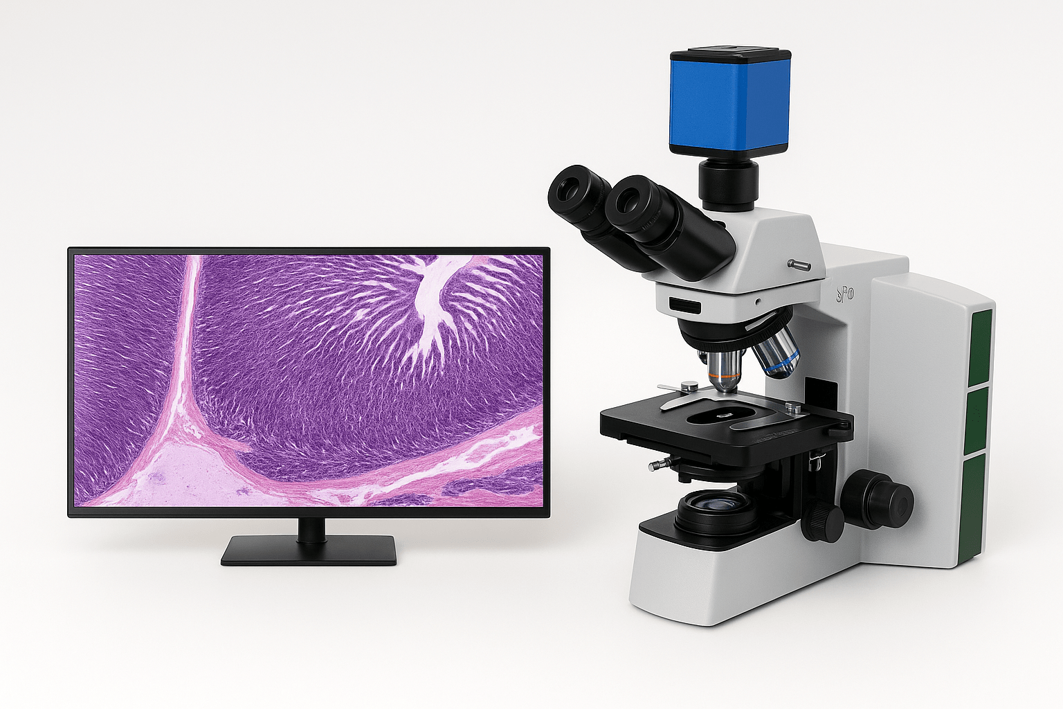
01 / Therapy guide
Cell therapies at a glance
A guide to established treatments and early-stage research. Select “Details” for more context. Status labels are for reference, not treatment advice.
| Intervention | Health category | Category | How it works / target | Reference status | Details |
|---|---|---|---|---|---|
| OSK Factors (Oct4, Sox2, Klf4)Cellular reprogramming concept | Cellular | Gene / cellular | Cell-state reprogramming | Experimental | |
| Tet-On / Doxycycline SwitchInducible gene expression | Cellular | Gene regulation | Drug-responsive expression control | Construct-dependent | |
| AAV Gene-Delivery VectorsViral vector platform | Cellular | Gene delivery | Delivery of a designed genetic payload | Platform; indication-specific | |
| Ex Vivo Gene-Edited Cell Therapy (CRISPR/Cas9)Patient-derived, genome-edited cells | Cellular | Gene-edited cell therapy | Edits a selected gene target in cells outside the body before reinfusion | Product-specific approval | |
| VC6TF Chemical CocktailSmall-molecule reprogramming framework | Cellular | Chemical reprogramming | Multi-pathway / epigenetic modulation | Experimental | |
| Valproic Acid (V)Small molecule · C₈H₁₆O₂ | Cellular | Chemical reprogramming | Histone deacetylase (HDAC) modulation | Context-specific research | |
| CHIR-99021 (C)Small molecule · C₂₂H₁₈Cl₂N₈ | Cellular | Chemical reprogramming | GSK-3β inhibition / Wnt pathway | Experimental | |
| Repsox / E-616452 (6)Small molecule · C₁₇H₁₃N₅ | Cellular | Chemical reprogramming | TGF-β receptor pathway modulation | Experimental | |
| Tranylcypromine (T)Small molecule · C₉H₁₁N | Nervous | Chemical reprogramming | LSD1 / MAO inhibition | Experimental | |
| Forskolin (F)Small molecule · C₂₂H₃₄O₇ | Chemical reprogramming | Adenylate cyclase / cAMP signaling | Experimental | ||
| Alpha-Ketoglutarate (AKG)Metabolic cofactor · C₅H₆O₅ | Metabolic / epigenetic | α-KG-dependent dioxygenase pathways | Investigational context | ||
| Sodium ButyrateSmall molecule · C₄H₇NaO₂ | Chemical reprogramming | Short-chain fatty acid / HDAC modulation | Experimental | ||
| Vitamin C (Ascorbic Acid)Small molecule · C₆H₈O₆ | Nutrient | Metabolic / epigenetic | Redox support / enzyme cofactor | Context-dependent research | |
| Adoptive Cell Transfer (ACT)Cell therapy | Immune | Immunotherapy | TIL / immune-cell transfer | Application-dependent | |
| Tumor-Infiltrating Lymphocyte (TIL) TherapyAutologous tumor-derived immune cells | Immune | Immunotherapy | Selected tumor-reactive lymphocytes expanded for cell therapy | Product-specific approval | |
| Regulatory T-Cell (Treg) TherapyImmune-regulation cellular therapy | Immune | Immunotherapy | Regulatory T cells support control of immune activation | Product-specific approval | |
| Pancreatic Islet-Cell TransplantationDonor-derived insulin-producing cells | Cell therapy | Transplanted islet cells can restore endogenous insulin secretion in selected settings | Product-specific approval | ||
| CAR T-Cell TherapyEngineered cellular biologic | Immune | Immunotherapy | Engineered T-cell antigen recognition | Approved for specific uses | |
| TCR-Engineered T-Cell TherapyGene-modified cellular immunotherapy | Immune | TCR immunotherapy | Engineered T-cell receptors recognize selected peptide antigens presented by HLA | Approved for specific indications | |
| CAR-NK Cell TherapyEngineered cellular therapy | Immune | Gene / cellular | Engineered NK-cell targeting | Research / experimental | |
| Bone Marrow Aspirate Concentrate (BMAC)Cellular preparation | Regenerative | Regenerative | Autologous bone-marrow cell fraction | Indication-dependent | |
| Autologous Chondrocyte ImplantationSurgical cell therapy | Regenerative | Regenerative | Cartilage-cell implantation | Approved for specific uses | |
| Induced Pluripotent Stem Cells (iPSCs)Stem-cell research platform | Cellular | Regenerative | Pluripotent cell generation / differentiation | Research / indication-specific | |
| Hematopoietic Stem Cell Transplantation (HSCT)Blood-forming stem-cell transplantation | Cellular | Stem-cell transplantation | Replaces blood-forming stem cells to re-establish blood-cell production | Established for specific indications | |
| Mesenchymal Stromal / Stem CellsCellular therapy research | Regenerative | Regenerative | Paracrine signaling and tissue-response research | Evidence varies by product | |
| Exosome / Extracellular Vesicle ProductsCell-free biologic concept | Cellular | Regenerative | Intercellular signaling cargo | Investigational / product-specific |
Gene regulation
Transcription factors shape cell identity. OSK is an experimental research concept, not a self-directed rejuvenation method. OSK in the table ↗
Cell therapy lifecycle
Source, processing, testing and follow-up each matter. Approval of one product does not transfer to another cell type or indication. FDA product status ↗
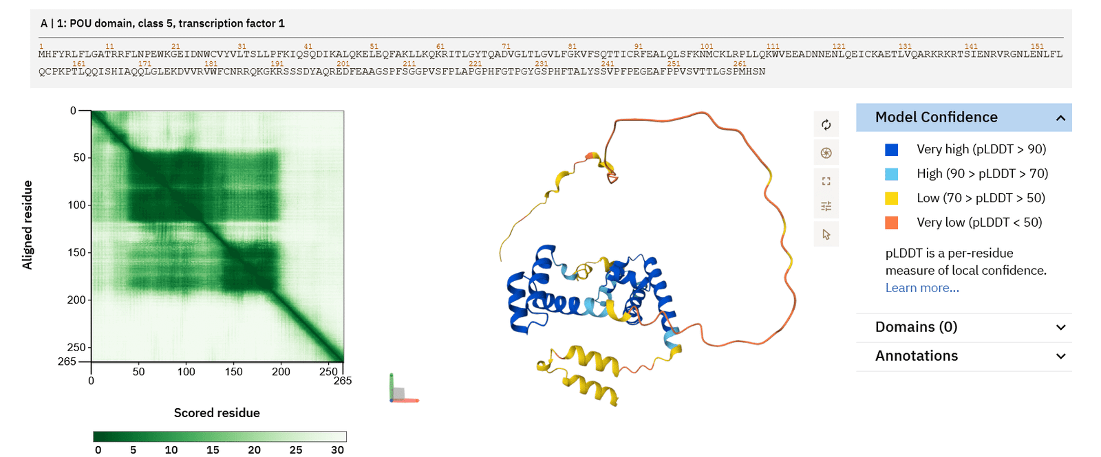
Protein structure context
Predicted and experimentally determined structures are different evidence types. Confidence scores do not establish function. AlphaFold ↗ PDBe / PDB ↗
How to review a cell-science claimEvidence checklist
Was it a biochemical assay, cultured cells, an animal model, a clinical study, or an authorized product label? Settings are not interchangeable.
Look for a prespecified endpoint, a meaningful comparator and a stated sample size. Mechanistic plausibility is not clinical benefit.
Cell products vary by source, processing and indication. Verify the exact product and its current, jurisdiction-specific status.
Check how adverse events, immune effects, persistence and long-term outcomes were monitored. Short windows can miss delayed risks.
02 / Microscope image review
What to look for in an image
Microscope images can be reviewed for cell shape, movement, and clusters. Results depend on image quality and careful human review.
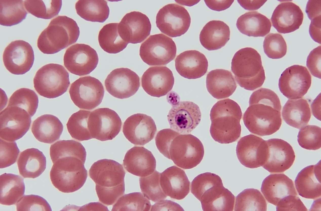
Compare the same microscope image.
Drag the divider right to compare the original Cell-Gem microscope image with a lightly sharpened view. Two red + markers and six small rings appear with the enhanced side and stay aligned; the key on the image explains each.
Display enhancement only. It does not add real microscope detail or diagnose disease. Any finding needs appropriate laboratory review.
What to look for
Common features to review
- 01SHAPE
Cell shape and size
Look at cell shape, edges, size differences, and visible changes across the image.
- 02MOTION
Movement
Follow movement and position changes over time, when the recording supports it.
- 03CLUSTERS
Cell clusters
Look for groups, chains, or coin-stack-like red-cell patterns. Appearance alone does not identify a cause.
- 04REVIEW
Other moving features
Changes in the fluid or background may need a closer look. Appearance alone cannot confirm a microorganism.
Video review tools
Computer-assisted review supports, but does not replace, expert checks
Track movement
Follow visible objects between frames and summarize movement or count changes.
Map cells and clusters
Group counts and locations into a consistent summary for review.
Flag unusual patterns
Highlight unusual shapes or clusters for a person to review, with further testing when needed.
Very small changes are difficult to measure. Results depend on the microscope, video quality, calibration, and a tested method.
The process
From image to review
Three steps from capture to confirmation.
- 1
Capture
Image cells with a microscope. Record sample context and check focus, lighting, and coverage.
- 2
Analyze
Software can track shape and movement, count cells, and flag unusual areas.
- 3
Review
A qualified reviewer checks flagged areas. Use validated methods for medical questions.
Review guide
Reviewing a microscope image
What to look at, how reported accuracy compares, and where review has limits. Results depend on image quality and qualified review.
What to look for
- Shape & sizeCell outline, size, and pale centers (central pallor) across the field.
- MovementPosition changes over time, where the recording supports it.
- ClustersGroups or chains of cells, such as coin-stack-like red-cell patterns. Appearance alone does not identify a cause.
- BackgroundChanges in the surrounding fluid. These cannot confirm a microorganism.
- Stain colorColor shows structure, such as a purple-stained nucleus. It does not identify disease.
Reported accuracy Unverified
| Metric | Manual | AI claim |
|---|---|---|
| Data extraction | 20–30% | 95–99% |
| Counting accuracy | ~75% | >98% |
| Diagnostic yield | 10–15% | 60–85% |
No citations, sample sizes, or test protocols were provided. These are not established benchmarks.
Limits of image review
- Not a diagnosisAI flags and cell appearance do not diagnose disease.
- Not an identificationAppearance alone cannot confirm a microorganism.
- Not a treatment guideImages do not show whether a therapy suits a person. Use qualified review and validated laboratory methods.